-
Tathagat Heart Line
080 22357777/41410099,
+91 9900356000 -
Bangalore
#31, Mallige premises,
Crescent road -
OPDs
09.00am - 06.00pm
Echocardiography is also referred as Echo, Doppler ultrasound of the heart, Cardiac Ultrasonography, 2D echo test, Cardiac Ultrasound, and 3D echo test. This test helps to represent the image of heart with the help of sound waves. It can be taken simultaneously with a stress test to determine the heart beat while the patient is doing exercise.
Echocardiography is a test that uses sound waves to show moving pictures of the size and shape of your heart. It also displays the working condition of your heart’s chambers and valves. Echo can also display zones of heart muscle that aren’t contracting well due to lesser blood flow or injury resulting from previous heart attack. A type of echo known as Doppler ultrasound displays the flow of blood through your heart’s chambers and valves.
Echo can detect imminent blood clots inside the heart, fluid buildup in the pericardium (the sac around the heart), and problems with the aorta. The aorta is the primary artery that carries oxygen-rich blood from your heart to rest of the body.
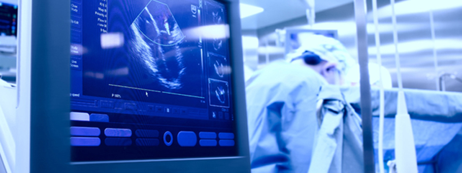
An echocardiogram (echo) is a test that uses high frequency sound waves (ultrasound) to make pictures of your heart’s chambers, valves, walls and the blood vessels (aorta, arteries, veins) attached to your heart. The test is also called echocardiography or diagnostic cardiac ultrasound.
- A probe called a transducer is passed over your chest. The probe produces sound waves that bounce off your heart and “echo” back to the probe. These waves are changed into pictures viewed on a video monitor.
- An echo can’t harm you.
- You can eat and drink before the test like you usually would.
The test helps to find out:
- The size and shape of your heart, and the size, thickness and movement of your heart’s walls.
- The heart’s pumping strength.
- If the heart valves are working correctly.
- If blood is leaking backwards through your heart valves (regurgitation).
- If the heart valves are too narrow (stenosis).
- If there is a tumor or infectious growth around your heart valves.
- Problems with the outer lining of heart (the pericardium).
- Problems with the large blood vessels that enter and leave the heart.
- Blood clots in the chambers of your heart.
- Abnormal holes between the chambers of the heart.